
Dr. Matthias Lütolf at TMM 2017
This year’s Till & McCulloch Meetings began with a rainy day on the beautiful mountainside in Mont-Tremblant, Quebec.
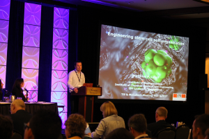
The morning talks started off with Dr. Matthias Lütolf. His lab at the École Polytechnique Fédérale de Lausanne, (EPFL) works on strategies to improve culture and growth of stem cells in lab settings. One of these strategies involves developing organoids. Organoids are basically mini-organs that the stem cells self-organize into after being exposed to predefined factors and structural microenvironments. The organoid structure has a similar cellular content, mechanical and functional feature to what is seen in vivo. Therefore, this mini-organ provides a great platform to study diseases and regeneration in tissues in a lab setting.
However, the common techniques used to generate organoids cause stochastic development and there is a huge distribution in the size and shape of the cells within them, which results in heterogeneous cultures. This is partly due to the fact that the cells in vitro typically do not have any spatial confinement or mechanical cues from the surrounding tissue. Dr. Lütolf’s group is trying to coax stem cells into forming a more robust and predictable organoid structure. They are exploring whether they can use bioengineering tricks to provide the missing environmental cues and signals by manipulating the 3D structure that the cells are cultured on.
To this end, they have tried to develop intestinal organoids in culture. Dr. Lütolf’s team has found that matrix mechanics and stiffness of the surrounding matrix plays an important role. A softer microenvironment would benefit intestinal crypt formation, and they were able to generate a fully defined microenvironment and mechanically guide the cluster of cells to form intestinal organoids.
They have also shown that geometry of the cellular environment can affect the patterning of the cells and direct gene expression. They were able to develop an open micro tissue system, shaped into a tubule, bearing crypt-like domains to resemble intestinal tissue. The stem cells appeared to populate the edges, and differentiated cells appeared at the villi of the microtissue.
Normally, an organoid culture is a closed cyst-like population of cells. With the open system, researchers will be able to modify the system and easily track the effects of those changes in the system. When the open microtissue was colonized by intestinal stem cells, the stem cells adhered to the tubular structures and generated colonies quickly, while also generating epithelial structures very similar to what is seen in organoid structures.
An organoid structure with predefined and well controlled mechanical and geometrical properties, and artificial signaling microenvironments like the one Lütolf’s group has developed, will give us valuable insight into tissue development. Also, these organoids can be used in drug development and regeneration treatment strategies.
Hamideh Emrani
Latest posts by Hamideh Emrani (see all)
- A second chance at life after a diagnosis of Multiple Sclerosis – Day 2 at TMM 2017 - November 16, 2017
- Taming the unruly immune system: the risky stem cell transplant that changed the fate of some Canadian MS patients – Day 2 at TMM 2017 - November 16, 2017
- Ingenious methods to engineer cells to treat diabetes – Day 1 TMM 2017 - November 9, 2017


Comments