
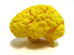
Image credit: Nevit Dilmen, NIH 3D Print Exchange, National Institutes of Health
Miniature human brains continue to develop when hooked up to a blood supply in the mouse brain in a finding that opens up new avenues for learning how the brain forms and falls ill, while at the same time raising ethical questions.
Last week, two teams reported success in transplanting primordial human brain tissue into mouse brains. The grafts survived thanks to nutrient flow via blood vessels that do not normally form when the neural tissue is grown in the dish.
Writing in Nature Biotechnology, a team led by Fred Gage, of The Salk Institute for Biological Studies in La Jolla, CA, described how the implants integrated into the mouse brains thanks to host blood vessels that supplied life-sustaining oxygen and nutrients. This allowed the grafts to survive for at least eight months, during which the human nerve fibers sprouted from them, made connections with the mouse neural circuits and exhibited neural activity.
Another team, led by Ben Waldau, of UC Davis Health, first co-cultured the mini brains with human blood vessels before implanting them into mice, as described in the journal NeuroReport.
It’s worth noting that the findings come on the 200th anniversary of Mary Shelley’s novel “Frankenstein; or, the Modern Prometheus” about a scientist who creates a monster in his lab by piecing together human and animal body parts. Similarities notwithstanding, by harnessing mouse skulls as living incubators, researchers hope to grow parts of human brains that are bigger and more complex than currently possible, for the purpose of shedding light on glitches that lead to a whole slew of brain disorders, from developmental (such as autism), to degenerative (such as Alzheimer’s).
When mini-brains, or brain organoids, were first described five years ago, they allowed scientists, for the first time, to peak into the early stages of the human brain as it formed in the dish. Grown from stem cells, these self-organizing balls of tissue strikingly resemble certain features of the fetal brain, including a distinct layered structure of the nascent cortex, which will take on cognitive functions.
Because they can be derived from patients’ stem cells, brain organoids are coveted for their potential to reveal clues about a person’s disorder and help find treatment best suited for each individual.
But there are caveats. Mini brains lack blood vessels and so their growth is sustained by nutrients from the culture medium. But these molecules have a hard time reaching the innermost cells after the organoids have grown to be about 4mm across – the length of a pencil tip. After a few months in culture, the mini brains start to die and shrivel.
The miniature brains also lack diverse immune cells that are a normal part of the brain circuit and whose miscues are thought to lead to mental disorders such as schizophrenia and depression.
Another limitation is that the organoids are separate entities and it is therefore not possible to study complex patterns of neural wiring that span different parts of the brain. Researchers in Austria made steps toward clearing this hurdle by stacking two organoids on top of each other, but the lack of blood supply remained a problem.
Building on earlier transplant research in rodents, the Salk team reasoned that it might also be possible to implant human brain organoids into mouse brains. They first grew the mini brains in the dish for 40 to 50 days before placing them into mouse brains from which some tissue had been removed to make room. The human cells were made to express a fluorescent protein that rendered them green and therefore easy to distinguish from the host cells. Observing the grafts through a tiny clear-covered opening in the skull, the researchers watched them thrive for the next eight months – the duration of the study – although the implants survived for longer.
A week after the transplant, mouse blood vessels started making inroads into the graft, carrying oxygen and food. The vasculature was essential for graft survival because implants with meagre blood supply shrivel away.
As the implants grew, they made more of mature cell types typically found in newborn brains, whereas the organoids of the same age, which continued growing in the dish, retained features of an early to mid-gestation fetal brain.
Two different types of immune cells also appeared.
After three months, nerve fibers extended from the graft forming information highways that carry the brain’s electrical activity. Crisscrossing the mouse brain, green human nerve fibers could be seen as far as the outer lobes of the opposite brain hemisphere.
Unlike the hallmark haphazard firing of nerve cells in cultured organoids, the implants had a synchronized neural activity as would be expected from a more mature tissue. Upon stimulation, the graft cells sent electrical impulses far and wide across the mouse brain, as detected by electrodes, suggesting that the graft was both physically and functionally integrated into the host nerve circuits.
The graft’s survival of 233 days is also an early proof of principle that mini brains can be used as transplant tissue to repair brain damage.
But, anyone thinking that mice with clumps of human brains inside their heads would turn out to be smarter is in for a disappointment. Mice with and without the implants looked and behaved the same, ruling out the possibility that the transplant procedure itself affected the experiments.
The Ethics
The study is the first published report of a human/mouse brain chimera and raises inevitable ethical questions. Can man-made human brain tissue develop enough to become sentient and what does this mean for future research? Previously, researchers reported the existence of photosensitive cells in cultured brain organoids. If the mini-brains can detect light, can they also feel pain? And does this become more likely as they grow bigger and more complex as part of the mouse brain?
No one is suggesting that the specks of human tissue from the Salk study imbued the much larger mouse brain with consciousness. But this was only the first step and, in the future, it may be possible to grow much bigger parts of the human brain. Could these larger and more complex structures contain glimpses of consciousness? And how would this be detected?
“Right now, we are able to grow small parts of the human brain – not all of it; but it may not be too long before we may be able to grow more,” says Liliana Attisano, Professor in the University of Toronto’s Department of Biochemistry, whose lab studies brain organoids. “The ethics of it is [sic] something we have to deal with,” she says.
Attisano said that overcoming the limitation of blood supply in organoid research is an important step forward. However, placing human tissue inside the confines of the skull does not make the study of human brain development any easier than when mice are used as stand-ins for humans, she said. The alternative approach of culturing vasculature and the miniature brains together could end up more promising for growing larger and more complex human brains that could be studied in the dish.
As the implanted or cultured human brains get bigger, so will the ethical concerns. And as with other issues in biomedical science—embryo-derived stem cells, gene editing—these will have to be weighed against the miniature brains’ potential to improve the lives of people with devastating disorders. Don’t count on implantable intelligence boosters any time soon.
Jovana Drinjakovic
Latest posts by Jovana Drinjakovic (see all)
- Canadian immunotherapy holds promise for patients with brain cancer - September 9, 2020
- Could stem cells be enlisted to battle COVID-19? - March 26, 2020
- Study reveals large differences at the molecular level between stem cells grown on different biomaterials - April 18, 2019


Comments