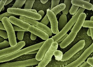
E. Coli bacteria. (Image by Gerd Altmann from Pixaby)
At first pass, the skin and the gut are not so different: both are lined with an outer layer of cells that serve as the first line of protection from biological, chemical and physical wear-and-tear. Accordingly, both the gut and skin are subject to tissue breakdown whether it be from invasive bacteria, rapid changes in pH, or physical injury. When it comes to treating the epithelial surface damage in the gut, however, the similarities between the gut and skin come to abrupt end.
Unlike our skin, where surface damage can be treated with a topical ointment and a run-of the mill bandage, the gut surface is coated with a thick layer of slippery mucous and located deep inside our body where a bandage would be difficult to apply and adhere. Moreover, the mucosal layer of the gut is constantly renewing itself, meaning that even the stickiest of bandages would slough off once the mucosal layer regenerates. Undeterred by these challenges, researchers from Harvard University have turned the problem on its head by designing a new type of “living bandage” that becomes stronger instead of weaker following mucosal regeneration.
This new regenerative technology was developed by researchers from the labs of Neel Joshi, PhD (with Anna Duraj-Thatte, PhD) and Jeff Karp, PhD (with Yuhan Lee, PhD) and appears in the August 2019 issue of Advanced Materials. The paper describes the living bandage technology as an injectable material made by, and composed of, Escherichia coli (E. coli) bacteria and their secreted polymers.
E. coli naturally generate large amounts of curli nanofibers, a type of self-assembling functional amyloid that is secreted during biofilm formation. The structural organization of curli nanofibers are highly versatile and have already been extensively investigated as biomaterial scaffolds that can be engineered at the nanoscale level. Their utility in regenerative medicine as a therapeutic, however, has been grossly underexplored – until now. Joshi and his colleagues discovered that when curli nanofibers were treated with a weak solution of sodium dodecyl sulfate detergent, they create a network that forms a thick and sticky hydrogel that is strikingly similar to the mucosal lining the gut epithelium.
The rheological properties of the hydrogel were further improved by engineering curli-trefoil factor fusion (TFF) protein domains, which are partially responsible for curli fibre cross-linking. The addition of different TFF domains enabled the fine-tuning of hydrogel mesh pore-size, endowing unique adhesive and physical properties suitable for an array of downstream therapeutic applications. Furthermore, engineering the E. Coli to secrete novel proteins fused to the curli nanofibers imparted adhesive specificity on the resulting hydrogels toward different areas of the gut. Whereas TFF decorated hydrogels preferentially stuck to the mucosal surface of a goat colon tissue sample, hydrogels displaying fibronectin-binding domains preferentially stuck to the fibronectin-rich serosal side of colon tissue samples.
Following engineered curli nanofiber production, the hydrogels were further filtered to produce either “live-gels” that contained E. coli or, instead, a cell-free alternative that was composed of the hydrogel alone. When orally administered to mice, the cell-laden live gels withstood the harsh environment of the gastrointestinal (GI) tract, accumulating in the cecum with the live bacteria intact.
Unlike the cell-free alternative, the live gel persisted following removal of the originally administered gel material during mucosal regeneration. The E. coli trapped within the live gels were able to continue producing protective curli fibers in the cecum, demonstrating the resilience and regenerative capacity of a single hydrogel dose. With respect to selective homing of the live gel to the cecum, Joshi adds: “The cecum is where E. coli most prefer to live, but we do think there’s a possibility that we could change the location [of the gel] in the GI tract by adding new proteins that bind to the upper GI tract or alternatively, employing different microbes that have preference for other parts of the GI tract.”
Joshi and collaborators hope to further develop this technology for use in fixing “holes” in the gut that develop during inflammatory bowel disease (IBD), a condition in which existing treatments have suboptimal outcomes.
On the therapeutic prospects of living bandage technology, Karp adds “with IBD, in general current drugs only work for a third of the patients and the drugs often stop working overtime so it is critical that we support continuous efforts to bring new therapeutics to bear for these patients. Living bandages could unlock a new paradigm for delivering therapeutics to the gut to address this pressing need.”
Joshi’s team also plans to improve their understanding of the mechanistic underpinnings of what makes these living bandages work so well. Looking forward, Joshi remarks: “There are questions to be answered of how the materials interact with the immune system of the gut. Moreover, self assembling functional amyloids are a common feature of microbial communities within the gut – our gut may be chock-full of these self-assembling proteins that could be useful in new applications.”
Living bandages represent a new approach in gut regeneration that chooses to work with, rather than against, the often hostile and hard-to-treat environment of the gastrointestinal system. By continuing to draw inspiration from how nature regenerates tissue, researchers may be able to harness the power of millions of years of evolution to create better treatments for patients in the 21st century. The Wyss Institute has put together a video of the living band technology that can be viewed here.
Erika Siren
Latest posts by Erika Siren (see all)
- Breathing new life into gut regeneration with living bandages - September 11, 2019
- The great experiment: CAR T in Canada - January 23, 2019


Comments