
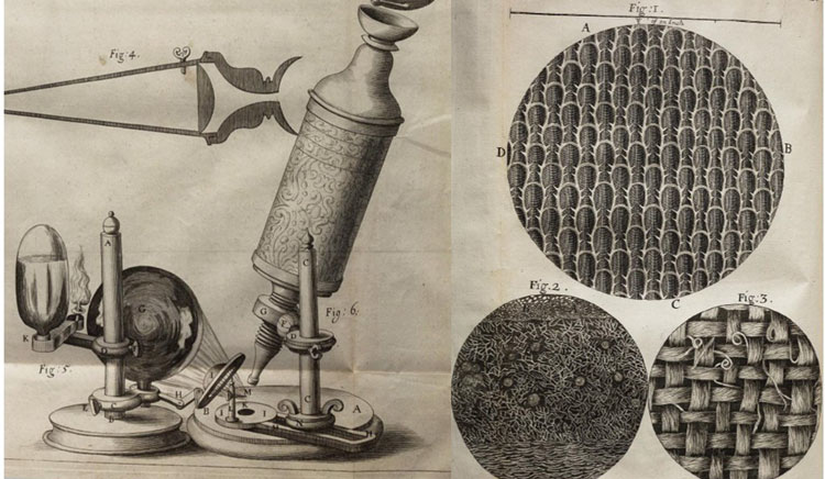
Left: Robert Hooke’s illustration of his microscope Right: surfaces of seaweed, rosemary and sage Micrographia, London, 1665
Since Robert Hooke first laid eyes on a micro-organism using his custom-built microscope in the 1660s, artists have deployed their skills in service of helping us to see the small picture.
Coining the word “cell” to describe what he saw through the lens, Hooke published Micrographia in 1665, complete with 38 incredibly detailed illustrations that were later turned into lavish copperplate engravings. Before embarking on his scientific career, Hooke had been an apprentice of royal court painter Sir Peter Lely (1618-1680), where he learned to draw and paint. Prefacing his book of illustrations, he declared that he had discovered “a new visible World.”
Our ability to peer into this “new visible world” has advanced quite a bit in the four centuries since. Fast-forwarding to 2008 (which already feels like a distant past given the pace of things), when Canada’s McMaster University announced it had installed “the most advanced and powerful electron microscope on the planet – so powerful it can probe the spaces between atoms.” “The resolution of the Titan 80-300 Cubed microscope is remarkable, the equivalent of the Hubble Telescope looking at the atomic level instead of at stars and galaxies,” said Dr. Gianluigi Botton, the project’s leader.
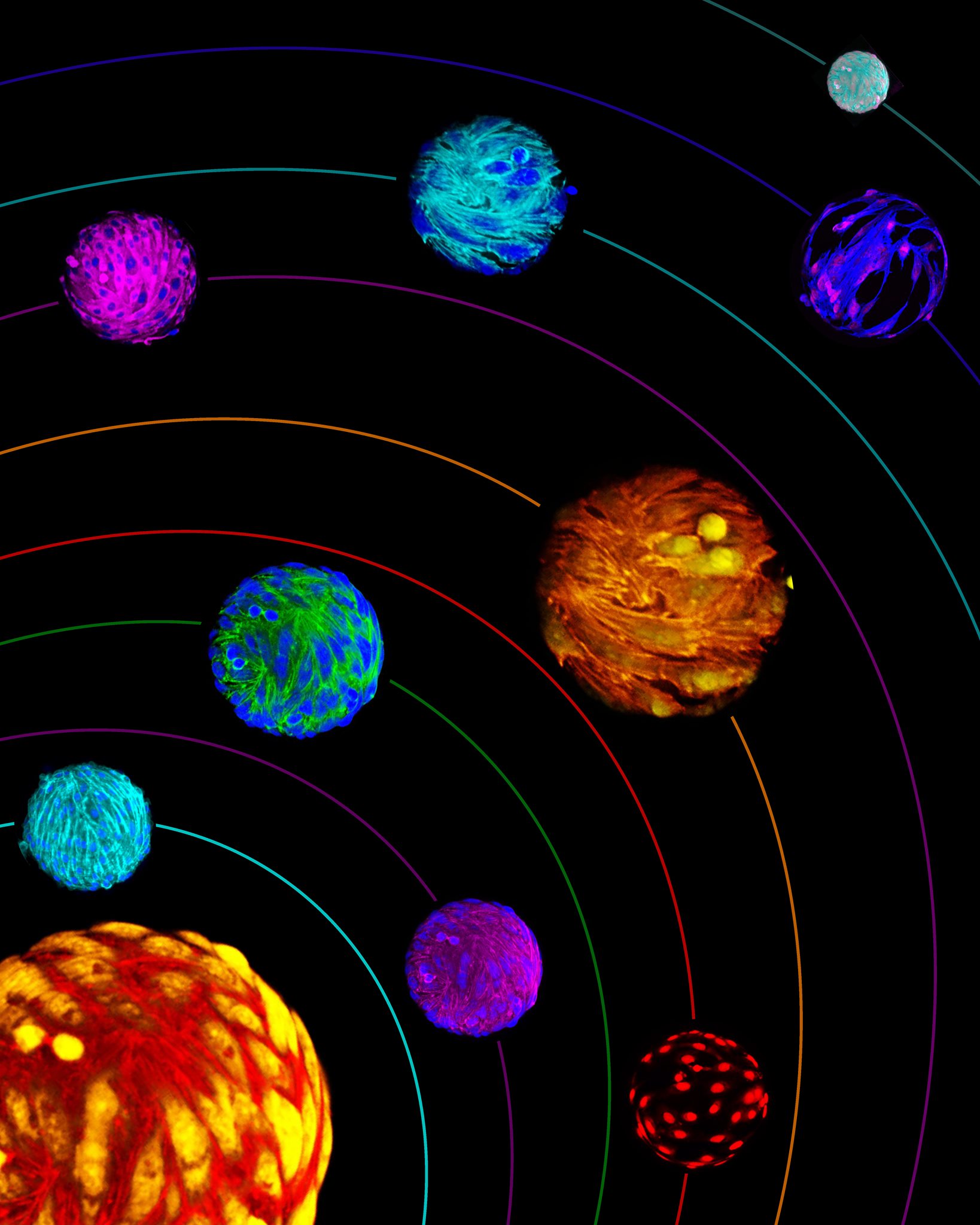
“Stem Cells Worlds of Possibilities” by Erin Roberts, University of Calgary, 2021
Evoking the cosmic to describe the microscopic has long been a recurring theme at the intersection of art and microscopy. Maybe the beauty of the macrocosm reflected in the microcosm has a way of capturing our imagination. I have a feeling that Erin Roberts, winner of the 2021 Cells I See art contest, might agree! “As above, so below,” the cryptic axiom with roots dating back to the eighth century invites us to contemplate.
Artistic portrayals of the microscopic have long broken free of their earlier, strictly representative function – as in Hooke’s Micrographia – giving us images bursting with flair, reimagined through the interpretive lens of artists.
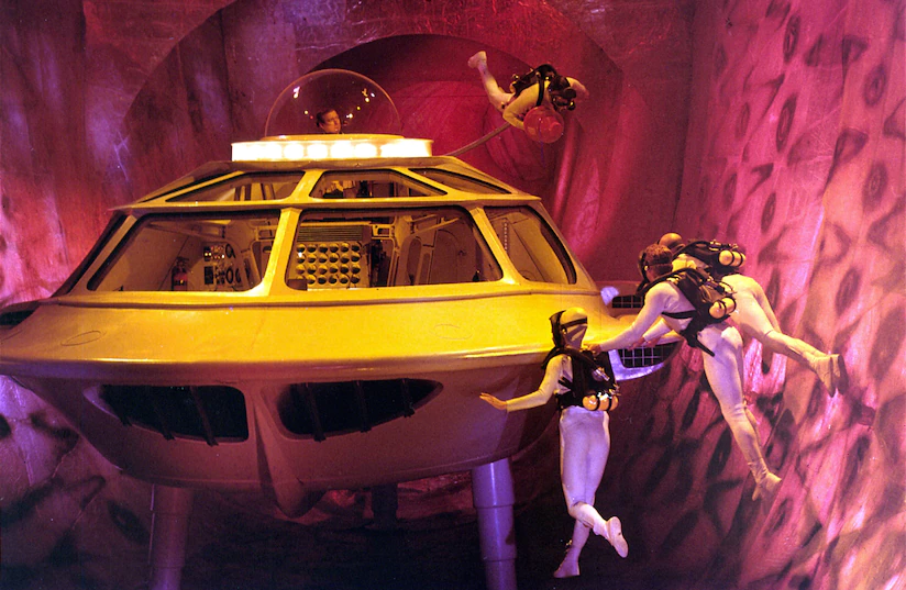
Credit: Turner Classic Movies, January 1996
The 1966 movie Fantastic Voyage imagined scientists who’d shrunk themselves in order to scuba dive inside a sick colleague’s bloodstream on a mission to heal him. “In one scene, antibodies attack a character in a wetsuit – like a school of predatory fish. The film assumed that the cellular world would be a miniature version of our own,” noted James Somers in his “A Journey to the Center of Our Cells” feature in the New Yorker this past February. Faced with the unknown, maybe we use art to fill in the gaps with what we do know – or think we know – about life on the human and cosmic scale.
But these gaps in our knowledge are being rapidly filled by teams of researchers, giving us a deeper understanding of how organisms behave, and in unprecedented detail. How will we capture and communicate this new, “new visible world”?
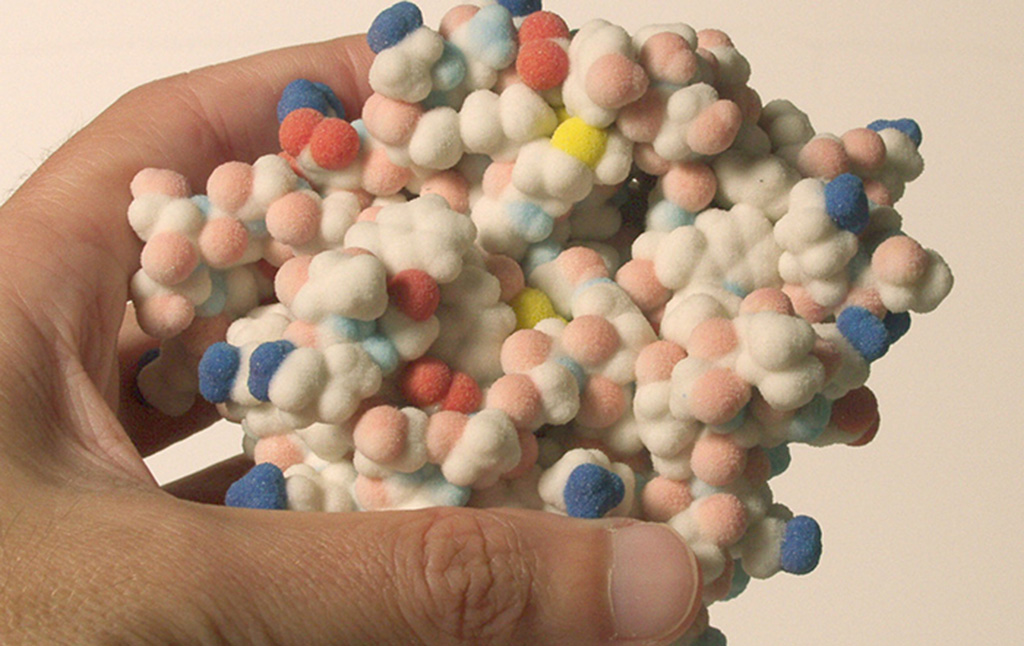
Credit: The Center for Computational Structural Biology (CCSB)
Teams working on the intersection of art and science are already on the case. CellPAINT-VR is an immersive, interactive virtual reality platform that visualizes molecular structures in complex cellular environments. James Somers, in his New Yorker feature, notes that Dr. David Goodsell of the CellPAINT-VR team is also a watercolourist and has had his works featured on the covers of the journals Cell and Nature.
Dr. Goodsell joins the ranks of past Cells I See art contest winners who have also been featured on magazine covers (as well as in galleries, science exhibits, calendars, and postcards.) If you have a flair for capturing the beauty of cells seen directly through the microscope or through the lens of your own artistic interpretation, why not enter this year’s Cells I See? Click here for the submission guidelines. Help us to see into the newest “new visible world” by submitting your entries by Tuesday, August 2, 2022. (Yes, there is prize money and bragging rights on offer.)
Cal Strode
Latest posts by Cal Strode (see all)
- What’s in the mix for 2026: ARM’s State of the Industry briefing - January 28, 2026
- World AIDS Day: Update on HIV cure research and gene therapies - December 1, 2025
- Headwinds and tailwinds for cell and gene therapy under the second Trump administration - March 11, 2025


Comments