
 Canada’s reputation for excellent science in the field of regenerative medicine can be traced back to the University of Toronto (U of T). Quite literally, Drs. James Till and Ernest McCulloch were U of T researchers when they discovered stem cells in the early 1960s. Not to minimize or dismiss excellent research happening across Canada – because it is equally impressive – but a substantial number of Canada’s world-class scientists are affiliated with this Toronto university.
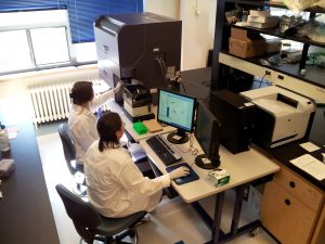
To get a sense of the regenerative medicine-based research happening at UofT, one only has to visit the Institute of Biomaterials and Biomedical Engineering (IBBME). CCRM is proud to work closely with IBBME’s researchers, to hire its graduates, and to share the exciting progress and developments coming from its labs.
The descriptions below are shared with permission of IBBME.
Rapid gene expression panel predicts effectiveness of treatments for leukemia patients
The standard treatment for patients diagnosed with acute myeloid leukemia (AML) is intensive chemotherapy, but patients vary widely in their response. Currently, it is difficult to predict who will do well with chemotherapy from those that would do better with novel therapies offered by clinical trials.
A team led by Stanley Ng, a senior PhD candidate in Professor Peter Zandstra’s lab, developed a new, rapid gene expression test. This test could help clinicians determine the best management for patients with AML by making it possible to accurately predict a patient’s response to chemotherapy within one to two days of diagnosis. The test is the result of a collaboration between the University of Toronto’s Faculties of Applied Science and Engineering and Medicine, the Princess Margaret Cancer Centre in Toronto, and leukemia clinics in France, Germany and the Netherlands.
In AML, leukemia stem cells (LSCs) are often resistant to standard chemotherapy. To develop the predictive test, Ng analyzed data produced by his collaborators, Drs. Jean Wang and John Dick at Princess Margaret, on genes more strongly expressed in LSCs. He identified 17 genes whose expression levels can be used to calculate a numerical risk score called LSC17 for each patient.
Ng then linked LSC17 scores with patient outcomes by gathering gene expression and clinical data from more than 900 AML patients treated at Princess Margaret and across Europe. His analysis revealed that patients with low LSC17 scores responded well to standard chemotherapy and survived longer than those with high scores. Princess Margaret is validating the test and plans to use it in a prospective clinical trial in several Ontario hospitals. The work is described in a study published in the journal Nature.
Exploring ways to stimulate damaged muscle to fix itself
If a patient suffers a debilitating muscle injury, a tissue graft often offers the best hope to restore lost function. Surgeons will replace the damaged muscle with healthy muscle from another part of the patient’s body or from a donor. However, the success of this procedure can be limited by the availability of grafts and the tendency for the new tissue to die before it incorporates into the patient’s body.
University Professor Michael Sefton and Professor Penney Gilbert are leading a multidisciplinary team that is taking a different approach, which could revolutionize how doctors treat skeletal muscle injuries. The researchers are exploring ways muscle stem cells can be stimulated to repair injured or damaged tissue, a process known as endogenous repair, instead of replacing injured tissue.
The team is one of 19 Medicine by Design-funded projects, announced on July 25, 2017, to accelerate discoveries in regenerative medicine and move them from the lab to patients more quickly. To get muscle tissue to fix itself, the researchers are testing whether a novel biomaterial scaffold can coax stem cells that are present in the muscle to spring into action to heal traumatic injuries. If successful, the team’s approach could shorten recovery, reduce pain and restore function sooner in patients around the world who suffer skeletal muscle injuries in car accidents, natural disasters and wars.
Sefton and Gilbert’s project brings together about 20 researchers from diverse fields, including clinicians, scientists and engineers who are experts in tissue engineering, stem cell biology and medicine.
Do physical stresses turn genes in muscle stem cells on and off?
Professor Penney Gilbert has been awarded a $1.4-million grant from the Human Frontier Science Program (HFSP) to lead a new, international collaboration to study how physical stress triggers the expression of genes in muscle stem cells.
While scientists have already uncovered many of the secrets that explain how and when genes are turned on and off, most of them involve ‘chemical cascades’ of signaling molecules and cell receptors. Many of these methods can take a long time to achieve the final result.
But Gilbert and her colleagues want to test the idea that under certain circumstances, physical forces—as opposed to chemical changes—could cause certain genes to become activated or deactivated.
The project, announced on March 27, 2017, enables Gilbert to combine her expertise in muscle stem cells with advanced methods in biophysics from Professor Timo Betz at the University of Münster in Germany and molecular imaging techniques developed by Professor Xavier Darzacq at the University of California, Berkeley. Together, the team will comprehensively examine the ways in which muscle stem cells transmit the physical stresses they experience into changes in their DNA and gene expression.
The results of the study could provide scientists with new, non-chemical strategies for turning genes on and off in muscle stem cells and possibly other types of cells. And, these findings could in turn help treat genetic diseases or other conditions caused when genes fail to turn on and off in the right place or at the right time.
Mending broken hearts—engineering a tiny tissue patch that could be injected rather than implanted
Repairing heart tissue destroyed by a heart attack or other medical conditions with regenerative cells or tissues usually requires invasive open-heart surgery. But Professor Milica Radisic and Miles Montgomery, a PhD candidate in Radisic’s lab, have developed a tiny patch of heart tissue with its own blood vessels that could be injected, rather than implanted.
After many attempts, Montgomery successfully designed a heart-repair patch with shape memory that unfolds into a bandage-like structure as it emerges from an injection needle. The injected patch unfolds to nearly the same size as a patch implanted by more invasive methods and the heart cells survive the procedure well. The researchers also showed that injecting the patch into rat hearts can improve cardiac function after a heart attack.
Radisic and her team are collaborating with researchers at the Hospital for Sick Children to assess the long-term stability of the patches and whether the improved cardiac function is maintained. The injectable patch could enable surgeons to use minimally-invasive techniques, which reduce recovery time, scarring and other risks. If the procedure could be performed successfully in human patients, it would significantly improve quality of life.
The researchers have applied for patents on the invention and are exploring whether the patch could be used in other organs, such as the liver. The research was published in the journal Nature Materials.
Transplanting healthy pancreatic cells under the skin to advance type 1 diabetes treatment
Researchers from University Professor Michael Sefton’s lab have demonstrated that the space under our skin might be an optimal location to treat type 1 diabetes (T1D).
PhD candidate Alexander Vlahos led a project that involved transplanting healthy pancreatic cells under the skin—an accessible and less hostile site—to produce insulin for blood glucose regulation. Once successfully implanted, these cells can then produce insulin to help regulate blood glucose levels. However, one challenge of using skin is that it has relatively few blood vessels.
Vlahos injected healthy pancreatic islets under the skin with a network support of blood vessels and found that normal blood sugar levels could be restored within 21 days, provided blood vessels are injected at the same time.
The next phase of their research will involve engineering the blood vessel network first with the hope that fewer islets will be required. This strategy would allow more of the cells to survive and function within the host, reducing the need for multiple donors per patient.
Vlahos’ study was published in the journal Proceedings of the National Academy of Sciences (PNAS). Earlier results of this work provided the basis for a $1.1-million research grant from international diabetes foundation JDRF to support a three-year study on this topic. The grant was announced on October 12, 2016.
New delivery strategies needed for more nanoparticle cancer drugs to reach their target
Targeting cancer cells for destruction while leaving healthy cells alone has been the promise of the field of cancer nanomedicine. But a new meta-analysis of 117 published papers, led by Professor Warren Chan and post-doctoral fellow Stefan Wilhelm, found that less than one per cent of designer nanoparticles reached their intended tumour target—and that figure hasn’t changed much over the last 10 years.
Their analysis showed that altering the nanoparticles to target cancer cells made little difference in net delivery efficiency. The majority of nanoparticles end up in the liver, spleen and kidneys, since the job of these organs is to clear foreign substances and toxins from the blood. This suggests researchers may have to control the interaction with these organs to prevent nanoparticles from being filtered out of the blood before they reach the target tumour.
One strategy Chan and Wilhelm pursued involved engineering nanoparticles that could dynamically respond to conditions in the body by altering their surfaces or other properties. This strategy may help the particles to avoid being removed by filtering organs, but have the optimal properties needed to enter tumours.
They also argued that a new systemic and coordinated long-term strategy is needed to increase nanoparticle delivery efficiency and that researchers will need to understand much more about the interaction between nanoparticles and the body’s various organs than they do today. To this end, Chan’s lab is also developing techniques to visualize these interactions across whole organs using 3D optical microscopy.
This particular work was published in the journal ACS Nano and can be found here. The full results of their review were published in Nature Reviews Materials.
To read more research summaries from IBBME, click here and here.
With files from the University of Toronto.
Stacey Johnson
Latest posts by Stacey Johnson (see all)
- Right Turn: Top 10 blogs from 2025 - January 9, 2026
- Right Turn: Season’s greetings and upcoming event - December 25, 2025
- Right Turn: Stem cell supplements: A growing market with growing risks - December 19, 2025


Comments