
Marissa Lithopoulos is a Ph.D. Candidate in Cellular and Molecular Medicine at the University of Ottawa. She works under the supervision of Dr. Bernard Thébaud at the Ottawa Hospital Research Institute. Marissa studies how brain stem cells are impaired in neonatal chronic lung disease and whether umbilical cord cells can be used to protect the lungs and the brain of preterm infants. Marissa is also dedicated to educating the public about stem cell biology and ethics. She has been the StemCellTalks Co-Chair for Ottawa and the National Advisory Committee. Marissa’s other passion is philosophy. She has been a teaching assistant for the Philosophy Department at the University of Ottawa since 2013, specializing in bioethics.
Spotted: 2017 Till & McCulloch Meetings in Mont-Tremblant
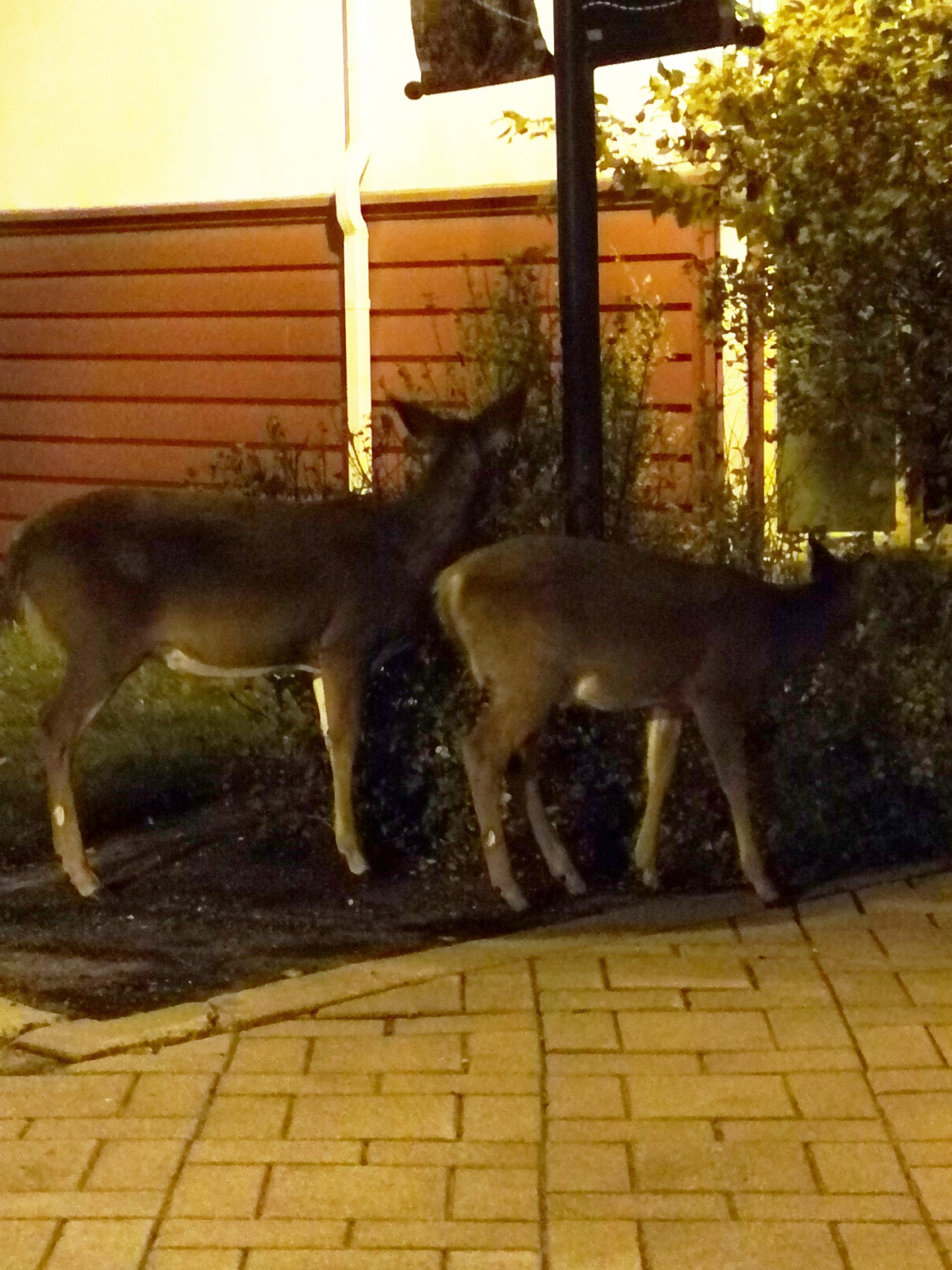
It’s that time of the year that brings joy to so many people. Breathing in the crisp air, reuniting with good friends, coming together from far and near to share in something much larger than ourselves. Oh, and if you are lucky, you might even see a reindeer or two along the way.
I am, of course, referring to Canada’s premier stem cell conference, the 2019 Till & McCulloch Meetings! It is an important meeting where scientists from all across the country exchange and share ideas to propel regenerative medicine forward and to strengthen Canadian stem cell research. This year’s presentations and talks showcased the impactful, exciting and unique research being conducted in Canada (and internationally too). Here’s a small sample.
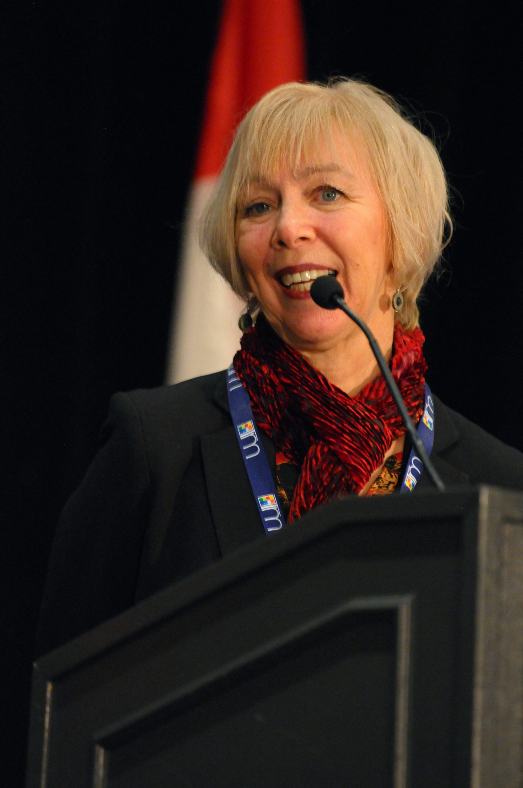
Dr. Freda Miller, Till & McCulloch Meetings Montreal, November 2019
2019 Till and McCulloch Award winner: Dr. Freda Miller, The Hospital for Sick Children
The Till and McCulloch Award honours the two Canadian pioneers of the stem cell field, Drs. James Till and Earnest McCulloch. At the conference each year, the prestigious award is presented to a stem cell researcher, based in Canada, who has made an outstanding scientific discovery. Dr. Freda Miller received the award for her peer-reviewed article, “Mesenchymal Precursor Cells in Adult Nerves Contribute to Mammalian Tissue Repair and Regeneration.”
Dr. Miller’s paper focuses on a novel mechanism by which peripheral nerves aid in tissue repair. The originality of the paper stemmed from its focus on the nerve mesenchymal cells, rather than on the neural components of the peripheral nerve.
Dr. Miller and her team found that after nerve injury in the digit tips of mice, mesenchymal cells migrate from the injured peripheral nerve to the neighbouring mesenchymal tissue. Then, based on environmental signals, the mesenchymal cells will repair the injured tissue by becoming the cell type that is required. For example, in the digit tip, if the skin is damaged, the mesenchymal cells can become skin cells and replace the damaged tissue.
This influential study highlights the importance of peripheral nerves for tissue repair and paves the way for harnessing the reparative potential of nerve mesenchymal cells.
Protecting our preemies: Dr. Bernard Thébaud, Ottawa Hospital Research Institute, University of Ottawa
Dr. Bernard Thébaud and his research team (myself included) are harnessing the reparative potential of another type of mesenchymal cell, those isolated from the umbilical cord, to help mitigate the complications of preterm birth.
Preterm birth is any birth that occurs before 37 weeks of pregnancy; however, babies born before 28 weeks of pregnancy (extremely preterm) are at the highest risk of life-threatening complications. Dr. Jen Gunter, an obstetrician-gynecologist and a mom of extremely preterm babies, describes the sad realities of preterm birth: “The legacy of extreme prematurity and the interventions that kept my boys alive left them with an assortment of medical conditions and disabilities, many of which I had never even heard of before.”
The most common complication of extreme prematurity is a chronic lung disease called bronchopulmonary dysplasia, or BPD. BPD is characterized by an arrest in lung growth, leaving these vulnerable neonatal patients with life-long breathing problems. These problems are caused by oxygen supplementation and mechanical ventilation, two life-saving therapies that unfortunately leave patients with many complications.
At the conference, Dr. Thébaud showcased his lab’s work to bring umbilical cord mesenchymal stromal cell therapy from the lab to patients. The pre-clinical experiments started with rodent models. Dr. Thébaud and his team showed that these mesenchymal cells can protect the lungs of rats that are exposed to supplemental oxygen (the same life-saving therapy that preterm human babies are exposed to, that leads to lung injury). This was an important first step to show the potential of these cells.
His lab then moved to a more clinically relevant model (i.e., one that mimicked humans much more closely). Dr. Thébaud and his team treated the world’s only preterm baboon model with their therapeutic umbilical cord mesenchymal stromal cells. Excitingly, they found that the cells were effective in helping the baboons survive the oxygen supplementation.
Dr. Thébaud‘s research shows great promise and this therapy is now moving to the clinic. Dr. Thébaud and his team are aiming to commence Phase 1 clinical trials next year, to assess safety of the cell therapy for preterm infants.
Reindeer to the rescue: Dr. Jeff Bernaskie, University of Calgary
Rudolph the reindeer, in addition to being Santa’s helper, may very well unlock the key to understanding how skin can repair itself without scar formation. Dr. Jeff Bernaskie and his team are studying the skin on the antlers of reindeer, called “velvet”. As Dr. Bernaskie states, “If we wound the velvet, it regenerates perfectly.” This is unique among mammalian species. Often in mammals, the skin forms a scar when repairing itself.
The novel study involved testing the reparative ability of the antler velvet compared to other areas on the animal. The researchers created small wounds on the antler velvet and the skin on the back of the animal (lumber region) and observed the healing over a period of 60 days. They found that the velvet had completely healed, with no observable difference from the surrounding skin, whereas the skin on the back of the animal formed a scar.
To determine whether the velvet could regenerate independently of the environment of the antler, the researchers grafted a piece of the velvet onto the back of the animal, then created a small wound in the velvet and again observed the healing over 60 days. Amazingly, even in this new environment, the velvet healed, scar-free.
Dr. Jeff Bernaskie and his team have introduced a novel animal model that acts as an ideal model for skin healing. Clearly, if researchers can better understand how the velvet regenerates, then they may eventually be able to develop new therapeutics to mimic this skin regeneration in humans.
Another year of research excellence in Canada
I am truly grateful to be a part of this Canadian research network, which provides tremendous support to trainees so that they can attend this exciting conference and meet scientists at various stages in their studies and research, from principal investigators to postdoctoral fellows to graduate students.
This event not only provides a great opportunity for networking and knowledge exchange, but also allows for forming great friendships with colleagues as well. One thing is very clear: It is through collaborations, discussion and shared resources that we will make great strides in regenerative medicine and that is the true strength of our Canadian based science. Here’s to another great year of stem cell research progress in Canada!
Guest
Latest posts by Guest (see all)
- Regenerative immunotherapy: Hope for chronic autoimmune diseases - September 16, 2025
- Canada’s regenerative revolution: Why AI is the catalyst - September 4, 2025
- Summer by Design: A launchpad for future entrepreneurs and industry scientists - August 14, 2025


Comments