
In this month’s installment of the “Bioprocess and Bioanalytics” blog, we get part II from Drs. Robin Turner and James Piret on Raman spectroscopy and how this method can be utilized to tell us new information about cell therapy products. Both are Professors in the Michael Smith Laboratories at the University of British Columbia. Dr. Robin Turner has a cross appointment in the Department of Electrical and Computer Engineering, and Dr. James Piret is in the Department of Chemical and Biological Engineering.
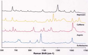
This image, while depicting a drug data set collected on a portable Raman system, offers an idea of what a Raman image could look like in cell manufacturing. (Source: Wikimedia Commons)
In Part 1 of this topic, we presented some of the theory and described the capabilities of Raman spectroscopy (RS). This is attracting attention as an emerging process analytical technology (PAT) for monitoring cell manufacturing processes. In Part 2, we will address practical questions that arise in such applications and comment on what the future may hold.
What does it take to implement Raman spectroscopy?
The utility of RS in other fields has driven the development of many commercially available components for custom configurations, as well as full stand-alone instruments with sophisticated software tools. Stand-alone systems vary widely in cost, from under $100,000 to more than $1,000,000 depending on the type of RS and desired features and accessories. Off the shelf, such systems are best suited to analyzing samples of cells or cell products that have been manually extracted from a process and prepared for exposure and analysis. In order to integrate automated RS measurements into a bioprocess, considerable customization or development by highly-trained and specialized personnel may be required.
Another factor that impacts cost is the choice of excitation wavelength, which in turn can limit the choice of instrumental platform and customizability. RS can be implemented with essentially any wavelength of excitation light from infrared to deep ultraviolet, with advantages to working in specific spectral regions for different applications. For most bioanalytical and biomedical applications involving cells, the near-infrared (NIR) region offers the best trade-off between sensitivity, susceptibility to fluorescence interference, and photodamage risk (directly or due to heating). Fortunately, this region of the spectrum is typically the least costly and easiest to work in, though it provides the lowest inherent sensitivity as elaborated below.
Why is Raman spectroscopy not more widely used?
All the desirable features of RS notwithstanding, there are challenges. The most restrictive being that Raman scattering is an extremely low-probability physical process. It is therefore not very sensitive, and therefore acquisition times from seconds to minutes per spectrum may be required to achieve satisfactory signal-to-noise ratios. Thus, high resolution Raman images comprising hundreds of spectral acquisitions can take hours and require the cells to be fixed. Different fixation methods have varied effects on the spectra and can complicate the analysis and interpretation. More advanced coherent RS methods (see below) can be much faster, but have their own shortcomings and are more costly. Fortunately, measurements averaged over a relatively large area/volume of live cells are more feasible and could be even more informative than single-cell measurements.
For any Raman application, the raw spectral data need to be processed to correct for signal intensity fluctuations (e.g. due to laser instability) and spurious background contributions from fluorescence or stray light. Nonetheless, artifacts can remain and the rich information carried by the spectra may be corrupted and misinterpreted. Even if all the information content is preserved with high fidelity, it can be challenging to extract the specific information of interest. This is mainly because RS is not inherently chemically selective. Rather, it detects particular vibrational modes (e.g. bending, stretching, ring breathing) that exist in a multitude of different molecules. For example, C-H stretching and bending modes are highly concentrated in lipids, but also present in other species. Observed vibrational bands can also be comprised of multiple overlapping peaks that may make interpretation difficult. There are other challenges, but most do not degrade the value and utility of a Raman spectrum as a label-free “fingerprint” measurement.
Outlook
Advances in laser and detector technologies, as well as new implementation strategies, are constantly improving the usefulness of RS and mitigating many of the challenges. For example, the low sensitivity of spontaneous Raman is not an issue at all for coherent anti-Stokes RS or stimulated RS that can even provide video frame-rate imaging of strong Raman bands. The needed instrumentation is costly and the specialized personnel are scarce, but this is changing.
In addition to improvements in instrumentation, the development of analytical methods suitable for RS is accelerating. New applications of multivariate statistical analyses are continuously adding to the “toolbox” of methods to identify spectral variations that are distinct from normal biological variations among or within samples. An exciting alternative to statistical methods is the prospect of applying new machine learning analytics to cell classification by Raman spectra. There are already impressive examples of this approach, currently focused mainly on medical diagnostic applications. Much effort is also being devoted to automation, efficiency and enhanced fidelity of spectral pre-processing operations. The output of these operations is crucial to the efficacy of any subsequent analytical steps. All of these software-based methods are also benefiting from improved hardware computational platforms.
For more than a decade, the FDA has been actively encouraging the testing of innovative PATs, even for monocultures producing a single protein, to improve the consistent manufacture of biologics. Bioprocess applications that exploit RS to monitor and control glucose levels are becoming established and, in the longer-term, we can expect increased applications to the monitoring of cellular therapy product quality. More information and a proof-of-concept publication where we applied RS to the analysis of embryonic stem cell differentiation is provided by Konorov et al. (87:10762−10769 Anal. Chem., 2015.
Sowmya Viswanathan
Latest posts by Sowmya Viswanathan (see all)
- Raman spectroscopy for monitoring therapeutic cell manufacturing – Part 2 - March 28, 2019
- Without new funding, key part of Canada’s stem cell research ecosystem to close - February 28, 2019
- Raman spectroscopy for monitoring therapeutic cell manufacturing – Part 1 - September 6, 2018


Comments