

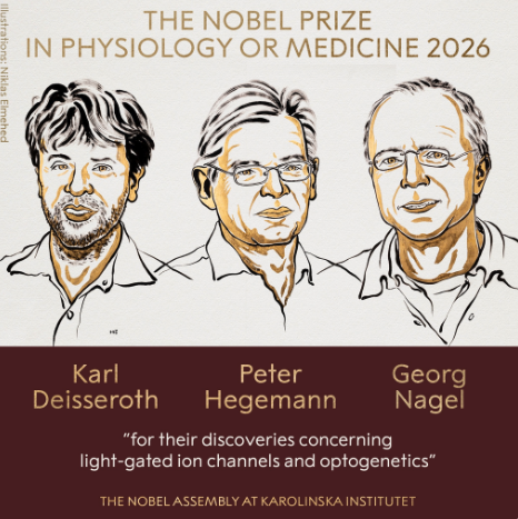
 Last week, optogenetics finally claimed the Nobel Prize in Physiology or Medicine. Most of the mainstream coverage thus far has placed a lot of emphasis on “turning parts of the brain on and off” in explaining the win; however, scientists had been doing that for years before optogenetics came along. The real reason why optogenetics took the Nobel this year goes much deeper. Here, I’ll explain what optogenetic tools do differently than their elder counterparts, and how optogenetics has found applications in other fields, because it’s no longer just neuroscience that’s benefiting. You’ll catch optogenetic tools in the fight against blindness, cardiology research, stem cell biology and more! Let’s jump right in.
A brief primer on opsins
The Nobel win began with green algae: single-celled photosynthetic organisms that detect light and swim toward it for food production. Green algae use a protein called channelrhodopsin (ChR) to do this. As the name implies, ChR proteins are ion channels that open when exposed to light of a certain wavelength. They work with a vitamin A-derived co-factor called all-trans-retinal (ATR), which is already found naturally in mammalian cells and doesn’t need to be supplied by researchers. This availability sets ChRs apart from previous, more complex attempts to control neurons with light, as awesome as they were. ATR changes shape when light hits it, and this causes the entire channel to open up. See the landmark paper on ChR1, and this one on ChR2. Two of the three Nobel winners – Nagel and Hegemann – discovered and characterized these proteins, landing them the prize.
ChR2 is a particularly important player in this story; when blue light hits ATR and the channel opens, sodium floods into the cell. In algal terms, this means it’s heading in the right direction. In neural terms, a flood of sodium into a cell is what allows it to fire electrical impulses. Recognizing this, Deisseroth (the third Nobel winner) engineered the gene for this protein into brain cells. The rest, of course, is history.
Since ChR2, many new opsins – the term we use for light-sensitive protein actuators – have either been discovered or artificially created in the lab. ChR2 was truly just the beginning. Why do we need so many? I explain below; but first, we have to discuss what makes optogenetics different from other modalities.
Life before optogenetics
Neuroscientists were already mapping neural circuits using electrical stimulation in humans and animals. Electrical stimulation is useful because it’s inherently the language of the brain, and it’s temporally precise. Electrical pulses are really quick. You can easily track behaviour as it relates to different types of stimulation patterns and durations. You can also see near-immediate differences between periods of time where stimulation is on versus when stimulation is off. For example, rats will enthusiastically press on a bar (over, and over, and over again) for electrical stimulation to their reward circuitry.
But there’s a problem with this approach: Each part of your brain is a mix of different neuronal cell types – GABA, dopamine, serotonin, and so forth. Electrical stimulation zaps them all at once, meaning it’s not helping to determine the functions and contributions of individual cell types. Neurotransmitter information is critical for drug development efforts. Also, if you want to see what happens when you turn neurons off, in most cases you’d have to create an electrical lesion. Given that lesions are a permanent burn made in the tissue, they’re tougher to accommodate in experimental designs and animal welfare, depending on the region of the brain. It’s also risky in humans for obvious reasons.
Parties more interested in specific, cell-type functions could instead use pharmacological manipulations, including receptor agonists and antagonists, or innovative chemogenetic tools. I recall an era of DREADDs, Designer Receptors Exclusively Activated by Designer Drugs. You could engineer your cell type of interest to express an entirely synthetic lock-and-key chemical system that could selectively activate or inhibit neuron classes without as many off-target effects.
The drawbacks of these approaches, however, are the timescale of action, in addition to the challenges with spatial selectivity. Pharmacological and chemogenetic approaches rely exclusively on brain injections of either a drug or an agent for genetic engineering. It’s tough to inject volumes limited to the brain structure of interest because liquids naturally diffuse as far as physics will take them. In addition, you can’t turn chemicals on and off in a temporally precise way to study activity patterns: Are the electrical impulses evenly spaced? Do they arrive in quick bursts? Are they variable? There’s no way to know.
It was simply down to a large, relatively unnatural flood of chemicals that either activate or suppress activity. You couldn’t turn it off unless you added another chemical to cancel it out again (and that also takes time); otherwise, you waited for the “washout” period, meaning the body would just metabolize and expel the chemical on its own. On the other hand, there were chemical toxins that could lesion an area and allow you to see what happens, but again the permanence of that is not ideal, and the damage could travel farther than intended.
So, in the lead-up to optogenetics, there was a trade-off. Researchers had to choose between controlling when they stimulated the brain and controlling what they stimulated. It’s a bit like making music with a piano: you could either strike groups of adjacent keys at exactly the right moments, or play individual notes without controlling the rhythm. Neither approach let you perform a complete melody.
The new era: Optogenetics, everywhere
Enter the era of ultraprecision. We can now engineer specific types of cells to respond to light; there’s nothing faster than that. Now we have both layers of control at once. You can figure out what neuronal cell type drives a given behaviour or function and what sort of coding language is being used. You can test different firing patterns (as I have!) and control their activity in a reversible, impermanent way. Another powerful improvement is spatial control. Even if your viral infusion for the initial genetic engineering travels farther than intended through the tissue, only the illuminated volume will be affected, and this is easily modelled. Some people are even illuminating just a few cells at a time with holographic optogenetics to try and reproduce specific local firing dynamics.
Earlier, I mentioned that it’s no longer just ChR2 that we have to work with. It was the first opsin protein used, but scientists have since discovered and synthetically created many more new variants to test out different hypotheses. Scientists studying neurons that fire very quickly are better off using ultrafast variants such as the ChETA – my personal favourite. Others require highly sensitive opsins that can be stimulated through the skull. Some users inhibit neurons at the cell body and go with a run-of-the-mill halorhodopsin, while others need inhibition at the axon terminal and choose an eOPN3 opsin from the humble mosquito.
As for matters of application, I’ve written about multiple optogenetic tools engineered to try and replace lost photoreceptor cells of the eye in degenerative eye diseases. This is possible because you have neurons in your eyes, and they allow you to see in cooperation with other front-line, light-sensing cells. If those light-sensing cells are lost, you can make the neurons themselves light-sensitive instead via optogenetic tools. Ongoing Phase I through III clinical trials thus far have yielded highly positive results, ranging from findings of safe outcomes to demonstrated restoration of functional vision.
That being said, optogenetics has now moved beyond the brain. Scientists are looking into how optogenetics can be used for severe, intractable pain conditions. Optogenetic tools are being used in ALS research at the interface of nerves and muscles. I’ve seen papers exploring the use of optogenetics to study cardiac musculature and heart regeneration. Stem cell biologists have used it in the study of differentiation. The field has absolutely exploded as a result of Deisseroth’s groundbreaking success in 2005.
Interestingly, teams are trying to get optogenetics to work in the human brain for therapeutic purposes. Right now, electrical deep brain stimulation (DBS) is approved by Health Canada and the FDA for multiple neurological disorders, including Parkinson’s and essential tremor, and it’s being researched for many more.
While it’s highly effective in its approved indications, the therapy is not perfect for the reasons I mentioned earlier: you can’t choose which brain chemical is affected in a manner that is as precise as optogenetics, though optimized patterning can help activate certain neuron cell types over others. In addition, different regions will react differently to the same electrical pulses, as they all have different properties and, importantly, different network inputs.
With optogenetics, treatments for neurological diseases could have much stronger specificity. Ensuring that toxicity and immunogenicity are brought under control remains the first priority. Also, human brain structures are larger than their counterparts in rodents, which means getting far more light into the brain (and the dangers of biological tissue heating do grow with light intensity). For this reason, it may be helpful to employ the specialized, highly sensitive opsins that I mentioned above – less light will be required for the tool to work, reducing heat-related side effects.
The last noteworthy element in need of optimization is delivery. Gene therapy in humans isn’t perfect yet, and for a prime example of this, see Sarepta’s recent issues with viral vector delivery and liver toxicity. However, it should be noted that what we learn in animal studies using optogenetics is directly informing DBS and other therapies already. For now, these data provide strong indirect benefits to various clinical interventions.
Long awaited
I hope that I’ve made the breadth and depth of significance associated with optogenetic tools a bit clearer. Though DREADDs and other innovative methodologies were critically useful and highly advanced, they could never quite solve enough problems all at once. As optogeneticists, we’ve been waiting for this moment for years because we experienced the symphonies ourselves, first-hand, and we’re in awe. Indeed, the team swept the awards that typically predict a Nobel. Does the technique still have some way to go before it reaches its full potential? Absolutely. However, the foundation is there, and the paradigm shift is complete.


Lyla El-Fayomi
Latest posts by Lyla El-Fayomi (see all)
- A Nobel for optogenetics: The real reason behind the win - October 19, 2026
- Optogenetic tools have officially changed the fight against blindness - June 24, 2026
- 2025 Recap: Regenerative Medicine News Under the Microscope - March 12, 2026


Comments