

Jainu Ajit is a freelance science writer based in Toronto. She holds a PhD in biomedical engineering from the University of Chicago, where she focused on improving vaccines and tumour therapeutics. She also completed a postdoctoral fellowship at Harvard Medical School, where she developed tools to access the blood-brain barrier. Jainu now works with startups and biotech companies to translate complex science into clear content.
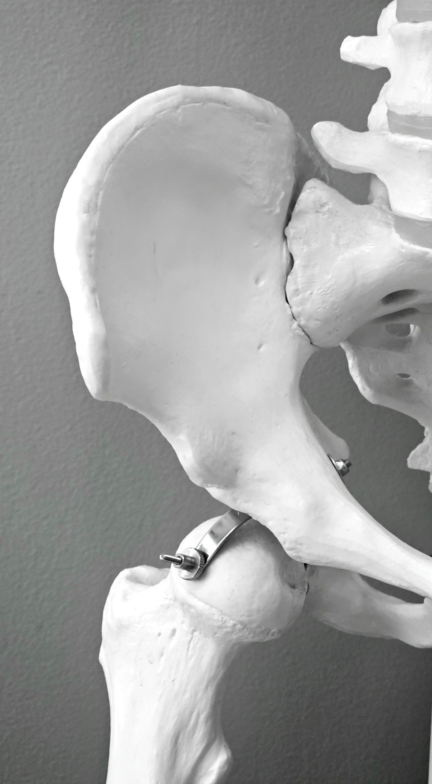
A hip bone. Image credit: Pexels.com
Osteoporosis weakens bones and increases fractures, particularly in menopausal women. In a Phase 1 study of 10 women with advanced osteoporosis, a single infusion of engineered bone marrow-derived mesenchymal stem cells (BM-MSCs) was found to be well tolerated with no treatment-related serious adverse events. The study also reported a significant reduction in fractures post-treatment, with effects lasting over five years.
Osteoporosis and menopause
Bone is constantly renewed by new cells through a process called bone remodelling. Aging can disrupt this balance, causing bone breakdown to outpace bone formation. Over time, this can lead to osteoporosis, a disease characterized by weaker bones and an increased risk of fractures.
Globally, osteoporosis is associated with one fragility fracture every three seconds. Women are particularly affected by osteoporosis after menopause. Menopausal women experience a loss of estrogen, a hormone that is crucial not only for their reproductive function but also for bone health. Estrogen keeps osteoclasts, cells that break down old and damaged bone, in check. As estrogen levels fall, this brake is released, leading to higher rates of osteoclast-mediated bone breakdown.
Although osteoporosis has no cure, anti-resorptives can lower fracture risk by inhibiting osteoclast activity. An alternate strategy is to boost osteoblast-mediated bone formation. These drugs are called osteoanabolics and include parathyroid hormone analogs.
The problem
A major limitation of existing osteoporosis treatments is the need for sustained treatment over time, along with associated side effects that worsen compliance. As a result, treatment adherence remains low, and a large proportion of patients discontinue treatment. Given that osteoporosis-related fragility fractures happen at a rate of 10 million every year, this gap needs to be addressed by finding an alternative that is both long-lasting and non-toxic.
Could BM-MSCs rebuild bone?
A new Phase 1 study by Moraleda and colleagues in Cell tested whether BM-MSCs could be safely engineered to home to bones in women with advanced osteoporosis.
MSCs have osteogenic potential and can be generated autologously. This reduces concerns of immune rejection compared with donor-derived cell therapies. In theory, improving their homing to bone could enhance their regenerative potential.
But there is a catch: native MSCs cannot find their way from the bloodstream to the bone marrow on their own. This is because they lack a ligand called sialylated Lewis X (sLeX), which binds to E-selectin, a molecule present on the blood vessels in the bone marrow.
To add sLeX to MSCs, Dr. Robert Sackstein, the senior corresponding author, previously developed a glycan engineering technique. The team noticed that MSCs already carried most of the sugar structure needed to make sLeX on a cell surface protein called CD44. The missing sugar was fucose. By adding fucose, they completed the sLeX motif on CD44. The team called this fucose-modified form of CD44 the hematopoietic cell E-/L-selectin ligand, or HCELL. HCELL acts as an address label, directing MSCs to bones by facilitating binding to E-selectin on blood vessels in the bone marrow.
The idea behind the modification makes sense, says Dr. Jeffrey Kiernan, HLA laboratory director and clinical investigator at UHN in Toronto, Canada, and Assistant Professor in Laboratory Medicine and Pathobiology at the University of Toronto, who was not involved in the study but has extensive experience in stem cell therapies.
Dr. Kiernan explained that the modification helps MSCs attach to and “roll” along blood vessel walls, an early step that allows cells to leave the bloodstream and reach the bone marrow efficiently.
The team later adapted the technique to produce clinical-grade fucosylated human BM MSCs that are suitable for use in patients. The reaction conditions were mild to cause minimal perturbation and preserve the genetic and functional properties of the cells.
Dr. Kiernan was particularly impressed by this careful progression from preclinical experiments to this first in-human study. For him, one of the study’s major strengths was this progression with years of stepwise work that established the framework that made clinical testing possible.
Study design
Preclinical experiments confirmed that exofucosylated BM-MSCs can home to bones to support bone formation. Additionally, since osteoporosis affects bones throughout the body, the team hypothesized that intravenous administration of exofucosylated BM-MSCs would be sufficient to induce bone formation. Using the patient’s own stem cells would reduce concerns surrounding adverse effects related to immune rejection compared to allogeneic therapies.
However, concerns about the safety of MSCs from older patients remained. In theory, these cells could accumulate genetic abnormalities that might increase the risk of malignancy.
This Phase 1 study therefore assessed the safety and feasibility of a single intravenous dose of exofucosylated BM-MSCs. The study tested two dose levels (2 million cells/kg and 5 million cells/kg) in 10 women aged 51-72 with advanced-stage osteoporosis and a history of fragility fractures. Most participants continued their existing osteoporosis treatments, including bisphosphonates or denosumab or the PTH analog teriparatide. The effects of exofucosylated BM-MSCs were evaluated alongside these standard therapies.
MSCs were harvested from the iliac crest (part of the hip bone), cultured in the lab, exofucosylated, and tested for sterility, fucose expression, and viability before the relevant doses were infused intravenously back into the patients. The researchers monitored patients for over five years after infusion.
Exofucosylated BM-MSCs: encouraging safety profile and fewer fractures
No infusion-related adverse events were observed in any participants post-infusion, supporting intravenous administration of exofucosylated BM-MSCs as a safe and feasible approach. No malignancies or tissue formations were observed during the five-year follow-up period.
To me, the most striking finding from the study was the marked reduction in fragility fractures. Compared with pre-infusion, fragility fractures decreased by 94 per cent two years post-infusion. Over the follow-up period, fragility fracture rates decreased from 0.54 to 0.11 fractures per patient-year after infusion. Mean bone tissue area and a measure of trabecular bone density also increased significantly after treatment.
How does a single infusion of BM-MSCs contribute to such long-lasting effects?
To explore how the treatment might affect bone remodelling, the team followed two key biomarkers at defined time points after infusion. (4,6,9,12,18, and 24 months)
- C-terminal telopeptide of type-1 collagen (b-CTx)- a marker of bone resorption generated when osteoclasts break down bone
- N-terminal propeptide of type-1 collagen (P1NP) – a marker of bone formation by osteoblast activity.
With just a single infusion of exofucosylated BM MSCs, levels of both these biomarkers increased. This simultaneous increase in markers of bone breakdown and bone formation indicates increased bone remodelling. However, the exact reason the effect lasts for years needs further mechanistic investigation, including biodistribution studies. Dr. Kiernan also noted that this type of mechanistic uncertainty is common in MSC research.
Conclusions
This Phase 1 study provides encouraging evidence that exofucosylated BM-MSCs can be administered safely and may produce durable osteoregenerative effects in patients with advanced-stage osteoporosis. A single intravenous infusion was followed by a long-lasting reduction in fracture incidence. However, establishing the treatment efficacy will require a larger randomized controlled trial.
Limitations
The small study size limited the generalizability of the findings. Mechanistic questions also remain about how a single infusion leads to long-lasting responses. The authors acknowledge the lack of biodistribution data to track the migration of BM-MSCs. Additionally, how a transient fucosylation step that lasts only 48 hours contributes to effects that persist for years remains unanswered.
From a promising technology to a scalable one
Another challenge with this therapy is manufacturing it at scale and cost-effectively. Dr. Kiernan sees the autologous nature of the therapy as a potential barrier to broader use. Manufacturing a separate cell product for each patient can be expensive and difficult to scale, he noted. An important next step, he says, would be to determine whether an allogeneic “off-the-shelf” MSC product could achieve similar effects without causing immune rejection. He also noted that exploring whether exofucosylation could improve the homing of other cell types could open new opportunities for cell therapy.

Guest
Latest posts by Guest (see all)
- Unlocking the regenerative potential of bone marrow stem cells in advanced osteoporosis - November 17, 2026
- Venture by Design: What it really takes to build viable companies in CGT - August 19, 2026
- Regenerative immunotherapy: Hope for chronic autoimmune diseases - September 16, 2025



Comments